Revealing How Hearts Develop in Humans
by Adrienne Mueller, PhD
September 22, 2023
Our hearts are complex organs made up of four chambers and a multitude of different cell types. Hearts are one of the first organs to develop in humans; with critical events occurring during the first three weeks of gestation. During mammalian development, the left and right ventricles of the heart arise, respectively, from patches of heart progenitor cells called the first and second heart fields.
Although the first and second heart field cell populations have been extensively studied in lab animals, finding them and studying them in live human tissue by working with human embryos has ethical and technical challenges. To address these challenges, researchers in the lab of Sean Wu, MD, PhD used human-induced pluripotent stem cells (hiPSCs) to recreate first and second heart field populations in vitro. Stem cells are a great means to model early human embryo development because of their ability to differentiate into many different kinds of embryonic cell types. However, until now, no lineage tracing tool has been able to follow the fates of progenitor cells to left ventricle and right ventricle heart muscles cells in human tissue.
In a study led by Francisco Galdos, PhD and recently reported in eLife, members of the Sean Wu lab and colleagues developed a novel cell lineage tracing reporter system that allowed the researchers to identify first heart field progenitors and their descendants – including left ventricle heart muscle cells. In addition, the investigators used the new technique of single cell RNA sequencing to profile the stem cell differentiation over time.
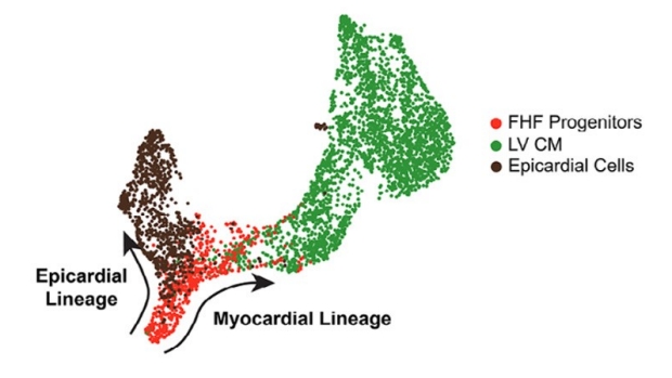
Depiction of first heart field development, showing the myocardial lineage first heart field progenitors (red), the epicardial lineage (brown), and left ventricle heart muscle cells (green).
Using the data from their reporter lines and single cell RNA sequencing experiments, the investigators discovered a predominance of first heart field cells differentiating into left ventricle heart muscle cells. Over 90% of left ventricular heart muscle cells in human stem cells were the progeny of first heart field cells. This is the first reporting of left ventricular-predomient differentiation of hiPSCs into heart muscle cells.
Galdos et al’s study provides the scientific community with a powerful new genetic lineage tracing approach, as well as a single-cell transcriptomic atlas of human stem cells undergoing cardiac differentiation. This study also shows that in vitro stem cells are capable of modeling early heart muscle cell development, which makes them a valuable tool for therapeutics and to better understand heart development disorders like congenital heart disease.
Additional Stanford Cardiovascular Institute-affiliated investigators who contributed to this study include Carissa Lee, Sharon Paige, William Goodyer, Sidra Xu, Tahmina Samad, Gabriela V Escobar, Aimee Beck, and Matthew H Porteus.
Dr. Francisco Galdos
Dr. Sean Wu