Novel Validation of Patient-Specific Blood Flow Model
by Micaela Harris
September 28, 2022
Cardiovascular diseases affect nearly half of US adults. Due to the prevalence of these diseases, accuracy, and reliability amongst cardiovascular imaging and modeling is critical. Specifically, modeling blood flow is a noninvasive means to pursue translational research. Computational fluid dynamics continues to gain popularity in personalized and predictive medicine, because computational modeling is fast, reliable, non-invasive, and low-cost. However, with these newly developed and ever-changing methods, it is crucial to validate and confirm that they produce accurate results. Specifically, it is important to validate cardiovascular blood flow models with techniques such as in vitro MRIs. MRI-based validation lets us confirm computational accuracy, which is necessary for us to continue to rely on these methods in the future. By using accurate computational models, procedures and processes could become less invasive and cardiovascular diagnosis and treatment may also improve due to the reliable nature of the techniques.
In an exciting new study led by Stanford University’s Ingrid S. Lan, PhD and Alison Marsden, PhD, investigators were able to achieve a novel corroboration of a sophisticated computational blood flow model with an in vitro patient-specific model to compare fluid structure interaction results. The investigators compared their computational model of fluid structure interaction with a constructed 3D model of a 50-year-old male subject’s “phantom” thoracic aorta. The researchers 3D-printed a phantom model to recreate patient-specific and life-like vascular structure and produce a simulation that was similar to the body’s processes. In order to simulate in vivo-like qualities, they also created an environment that mimicked blood density with a glycerol and water mixture, and pinch valves to recreate vascular resistance. The investigators measured the pressures created in the phantom model’s vasculature and compared it to their computational model for numerical accuracy.
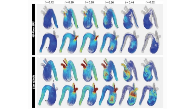
The image depicts correspondence between the 4D MRI of the phantom model (above) with the simulated computational blood flow model (below). Please refer to the article for the full image showing the computerized aorta model.
The investigators found strong agreement in comparing MRI scans of simulated blood flow in the 3D-printed phantom model with blood flow predictions of their computational model – demonstrating the validity of the computational model. By validating these numerical methods, this study provides strong support for using computational models as noninvasive and dynamic methods to produce patient-specific blood flow simulations.
Additional Stanford Cardiovascular Institute-affiliated investigators who contributed to this study include Weiguang Yan, PhD; and Daniel B. Ennis, PhD.

Alison Marsden, PhD