Treating Heart Failure from the Outside In
by Adrienne Mueller, PhD
April 22, 2021
When Heart Muscle Cells Die
Heart disease is the leading cause of death in the United States. Heart attacks and heart failure cause damage to heart tissue, which in turn causes heart dysfunction. Survivors of heart attacks are at increased risk for complications and are also often obliged to seek chronic treatment or device therapy. Because human heart muscle cells do not regenerate, once you lose them, they are lost forever. However, a new therapy is on the horizon that could help repair heart muscle cells that are damaged.
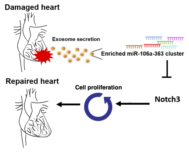
Damaged heart cells release exosomes that are enriched with microRNA cluster miR-106a-363, which then acts through the Notch3 pathway to increase cell proliferation and help repair the damaged tissue.
Extracellular Vesicles to the Rescue
Previous research from the lab of Phillip C. Yang, MD and others has shown that extracellular vesicles, small bundles of signaling molecules that are present outside the cell, can help repair heart muscle cells. However, in order to develop a treatment that would deploy these extracellular vesicles, or EVs, it is important to know exactly which molecules in the EVs are helping with the repair, and how they are doing so. A recent study by first author Ji-Hye Jung, PhD and senior author Philip C. Yang, MD sought to answer specifically these questions.
In their study, they compared the EVs of healthy tissue with tissue that has been oxygen-deprived. They hypothesized that the EVs produced by oxygen-deprived heart muscle cells would be more able to help heart cells regenerate than control EVs. As Jung et al report in their recent Basic Research in Cardiology publication, their hypothesis was correct. EVs from oxygen-derived heart muscle cells were better able to improve heart tissue viability and function both in vitro and in vivo by triggering the heart muscle cells to divide and proliferate.
Notch3: The Next Piece of the Puzzle
Jung et al then went on to investigate exactly which molecules were enriched in the EVs from the oxygen-deprived tissue. The investigators found that the EVs contained higher levels of a specific microRNA cluster (miR-106a-363). MicroRNAs are short, noncoding segments of RNA, and a microRNA cluster is several smaller microRNAs that are generated from one long, original long microRNA. Jung et al next demonstrated that this specific microRNA cluster alone was effective at preserving heart muscle cell function and reducing tissue scarring in the injured heart. They then went to identify that a key player in the signaling cascade initiated by this microRNA cluster is the Notch3 pathway. The investigators therefore show not only which microRNA causes heart muscle growth and repair, but that it does so using specifically the Notch3 pathway to promote heart muscle cells to proliferate.
This Yang lab study shows that a microRNA cluster never before associated with cardiovascular disease contributes to heart tissue repair through heart muscle cell division. Future heart failure therapies should consider targeting this microRNA cluster and its associated Notch3 signaling pathway to stimulate heart muscle cell regeneration and improve patient outcomes.
Additional Stanford Cardiovascular Institute-affiliated investigators who contributed to this work include Gentaro Ikeda, Yuko Tada, Daniel von Bornstädt, Michelle R. Santoso, Christine Wahlquist, Siyeon Rhee, Anthony C. Yu, Connor G. O’brien, Kristy Red‑Horse, Eric A. Appel, Mark Mercola, and Joseph Woo.

Ji-Hye Jung, PhD

Phillip C. Yang, MD