How mutations in a single gene can prematurely
age heart muscle cells
by Adrienne Mueller, PhD
December 15, 2021
Duchenne muscular dystrophy (DMD) is a rare but deadly disease that affects approximately 1 in 5000 boys. DMD causes severe muscle degeneration that eventually leads to heart and lung failure, and patients with DMD usually die before the age of 30. To date, there is no specific treatment for DMD: patients are reliant on non-specific heart failure treatments that do not tackle the underlying disorder caused by the lack of dystrophin, a protein that helps muscle cell contraction. The lack of specific treatment is mainly due to our not yet having a method to deliver or compensate for the missing dystrophin, or to combat the premature loss of heart muscle cells.
Previously, the lab of Helen Blau, PhD identified something unusual about the heart muscle cells of patients with DMD: they have shorter telomeres. Telomeres are DNA regions that cap and protect the ends of our chromosomes. As we age, telomeres shorten every time our cells replenish themselves by dividing. Telomere-shortening is a natural process for many of the cells in our body, but it is not natural for heart muscle cells which are one of the few cell types that do not divide. Therefore, in muscle cells of DMD hearts, telomere-shortening is initiated by an aberrant process that is not part of the cells’ natural cycle of aging.
The Blau lab sought to identify the mechanism behind this uncharacteristic telomere-shortening in DMD patients. To do so, they used human induced pluripotent stem cell-derived heart muscle cells of both healthy individuals and patients with DMD. By growing patient-derived heart muscle cells in a dish, the Blau lab was able to study what is different about the telomeres of healthy individuals and DMD patients without experimenting on the patients directly. Their study, led by first author Alex C. Y. Chang, PhD, and senior author Helen Blau, PhD, was recently published in Stem Cell Reports.
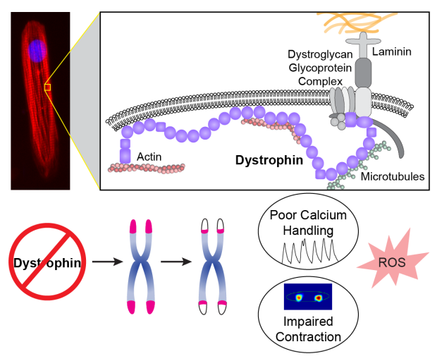
Dystrophin is a key component of heart muscle cells' cellular architecture. The absence of dystrophin causes shortening of chromosome-capping telomeres, poor calcium handling, impaired contraction, and an increase in reactive oxygen species.
Chang et al showed that, in the stiff tissue environment of unhealthy hearts, DMD heart muscle cells have several deficits -- they produce weaker contractions and have higher levels of reactive oxygen species – which are damaging to cells. The study also found an ongoing shortening of the telomeres in the DMD heart muscle cells that was related to a decrease in levels of protective, telomere-capping proteins called the “shelterin complex.” Chang et al. propose that when heart muscle cells attempt to contract in a stiff environment without dystrophin, the gene that is defunct in DMD, it is very stressful for the cells and produces reactive oxygen species.
Those reactive oxygen species can disrupt the binding of the shelterin complex to telomeres, reducing their protection of the telomere ends. Reactive oxygen species may even directly attack telomeres and contribute to their unexpected shortening.
In summary, contraction in a stiff heart tissue without the presence of the key structural contractile protein dystrophin is causally linked to telomere-shortening and the triggering of a senescence-like state in DMD heart muscle cells. New therapies should consider avenues that help heart muscle cells protect against telomere shortening.
Additional Stanford Cardiovascular Institute-affiliated authors who contributed to this study include Gaspard Pardon, Andrew C. H. Chang, Haodi Wu, Sang-Ging Ong, Asuka Eguchi, Sara Ancel, Colin Holbrook, John Ramunas, Alexandre J. S. Ribeiro, Kassie Koleckar, Joseph C. Wu, and Beth L. Pruitt.

Alex C. Y. Chang, PhD

Helen Blau, PhD