Linking Sugar Metabolism to Cell Proliferation
in Pulmonary Arterial Hypertension
by Adrienne Mueller, PhD
April 27, 2021
Too Many Cells
Pulmonary arterial hypertension (PAH) is a severe and chronic disease that worsens over time. In PAH, the blood vessels of the lung progressively narrow, and some become completely occluded. When this happens, PAH becomes life-threatening and as there is no treatment that reverses this process. Although the cause of PAH is poorly understood, we do know that individuals with PAH have abnormal growth of the smooth muscle cells of the lung blood vessels reminiscent of cancer. Abnormal growth of smooth muscle cells is responsible for the narrowing, and in some cases complete blockage, of lung blood vessels. Blood can only flow through the remaining vessels that have narrow openings if the pressure is very high. Because the side of the heart pumping blood to the lungs must maintain a higher and higher pressure, the heart muscle eventually weakens and fails. Several therapies currently exist to try to keep the lung vessels open as long as possible. These therapies improve quality of life and survival for a time, but eventually become less effective as the vessels become narrower or disappear.
Too Much Energy
The challenge therefore is to understand what is causing abnormal proliferation of lung blood vessel smooth muscle cells, and then to determine how to reverse it. Previous studies have shown that abnormal cell growth is associated with increased sugar metabolism – a means for our cells to produce energy. Suppressing sugar metabolism in lung smooth muscle cells, or cancer cells for that matter, can reduce cell proliferation by depriving the cells of the energy they need to divide. While it is known that cells that are rapidly dividing have intense energy demands, products of metabolism or metabolites can also drive changes in genes and proteins that may be necessary for growth. A study recently published in Circulation and led by first authors Dan Li, PhD and Ning-Yi Shao, PhD, and by senior author Marlene Rabinovitch, MD, found a single enzyme that connected energy demands with the genes required for rapid growth of smooth muscle cells in PAH.
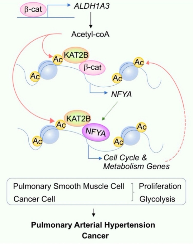
ALDH1A3 has a dual role in producing energy and in modifying chromatin at specific sites regulated by the transcription factor NFY to increase cell cycle genes that coordinate rapid division of cells and other genes that drive metabolism.
Relieving the Pressure
Li and Shao et al showed that smooth muscle cells of individuals with PAH exhibit an overabundance of the sugar-metabolizing protein ALDH1A3, an aldehyde dehydrogenase. The investigators then demonstrated that this specific sugar-metabolizing protein provides the cell with energy and causes epigenetic changes, or alters the landscape of the genome, to influence expression of genes that drive cell proliferation. ALDH1A3 is responsible for the production of a master transcription factor called NFY that increases expression of multiple genes required to coordinate rapid division of cells. When the investigators reduced the amount of ALDH1A3 in the smooth muscle cells, pulmonary hypertension was prevented because the vessels did not narrow with excessive smooth muscle cell growth. While reducing ALDH1A3 in smooth muscle cells had a beneficial effect on the development of pulmonary hypertension, the authors also observed that reducing this enzyme in the endothelial cells lining the blood vessel could have an adverse effect. This is an important consideration in developing a treatment that will target ALDH1A3 only in smooth muscle cells.
The Rabinovitch Lab investigators have therefore identified the specific mechanism underlying lung blood vessel narrowing in PAH: excessive activity of the sugar-metabolizing protein ALDH1A3 leads to the activation of a master transcription factor that increases expression of genes required for excessive lung smooth muscle cell proliferation and produces the energy the cells need to divide. ALDH1A3 is therefore a potential drug target in PAH, if it can be delivered to the smooth muscle cells of lung circulation.
Additional Stanford Cardiovascular Institute-affiliated investigators who contributed to this study include Jan-Renier Moonen, Zhixin Zhao, Minyi Shi, Shoichiro Otsuki, Lingli Wang, Tiffany Nguyen, Elaine Yan, David P. Marciano, Kévin Contrepois, Caiyun G. Li, Joseph C. Wu, and Michael P. Snyder.

Dr. Dan Li

Dr. Ning-Yi Shao

Dr. Marlene Rabinovitch