Stem cell-derived cardiomyocyte tool could improve drug safety
By Amanda J. Chase, Ph.D.
March 21, 2019
Stanford researchers have developed a tool to isolate specific subpopulations of cardiovascular cells to provide a model for more precise drug testing.
In today’s world, we are routinely reminded to exercise, eat healthy, and avoid tobacco use to decrease the risk of cardiovascular disease (CVD)s. Heart disease remains the leading cause of death for both men and women of most racial/ethnic groups in the US, with about 1 in every 4 deaths caused by CVD, according to the Center for Disease Control (CDC). In addition to lifestyle choices, there are other factors outside of our control that also contribute to the risk of being at-risk for CVD, such as inherited genes and other environmental factors.
The need to understand this combination of genes, lifestyle, and environment drives researchers to look for the study and development of new ways to treat and cure CVDs. The ability to study how these factors contribute to CVD was significantly improved by the development of patient-derived cardiac muscle cells known as cardiomyocytes (CMs). Researchers can collect cells from the blood of patients, and then manipulate treat them such that they can become nearly any tissue in the body. When those cells are turned into heart muscle cells, called cardiomyocytes, they faithfully mirror the expression of specific genes in the donor’s native heart tissue. Therefore, these cells provide a platform for better drug screening and disease modeling, as well as and the first step toward realizing the beginnings of a more personalized approach to studying and treating heart disease. Furthermore, these patient-derived CMs (called induced pluripotent stem cell-derived CMs; iPSC-CMs) can be used to generate engineered heart tissue (EHT) to better mimic the complex environment of a living human heart. Within the developing heart, there are ventricular, atrial, and nodal CMs; the ventricular and atrial CMs form the muscle walls of the heart (myocardium), whereas the nodal CMs function to set and maintain the rhythm, or beating pattern, of the heart. Having a highly homogeneous pool of ventricular or atrial CMs would be beneficial for applications such as determining drug safety, cardiac evaluation, and cell therapy for heart attacks by allowing personalized cardiac repair. A team of researchers primarily affiliated with the Stanford Cardiovascular Institute sought to find a way to make the heterogenous iPSC-CMs into more homogenous population of cells. Led by first author Joe Z. Zhang, Ph.D., and senior author Joseph W. Wu, M.D., Ph.D., Director of Stanford Cardiovascular Institute, they developed an innovative tool to helps isolate specific cardiac subpopulations to generate a purer population of cells. The work was published in Cell Stem Cell.
Heart development is orchestrated by the regulated expression of well-defined cardiac factors. Two important factors, called TBX5 and NKX2-5, control aspects of heart development in a certain part of the heart during particular and at certain times. In this study, researchers developed iPSC-CMs that expressed TBX5 and/or NKX2-5 with a colored label to help researchers identify cells that expressed both factors, either factor, or neither factor. Using this tool, they were able to isolate four different subpopulations. “Cell identity is determined by the expression of genetic markers,” explains first author Dr. Joe Z. Zhang, a postdoctoral fellow in Dr. Wu’s lab. “We first measured the gene expression of individual isolated subpopulations, and we found that each subpopulation has a unique pattern of gene expression, indicative of different cardiac lineages. We then performed a set of functional studies to confirm that the four subpopulations belong to different cardiac lineages.” Taking the study further, researchers also found a marker (CORIN) that was expressed in only one subpopulation of CMs (ventricular CMs), which allows for the isolation of those cells for preclinical studies.
The tool developed by these researchers allows, for the first time, isolation of lineage-specific cardiovascular cells. This is important because it will facilitate more precise testing of the potency and safety window of new drugs that target specific cardiomyocyte subtypes, improve disease modeling, and advance the tissue engineering field by generating pure ventricular EHTs. “The purified cardiac subpopulations provide a valuable platform for precise drug screening because different types of cells have a different response to the same drug at the same dose,” said Joseph Wu, MD, PhD, director of Stanford Cardiovascular Institute and Simon H. Stertzer, MD, Professor. Dr. Wu adds, “Our tool now allows researchers and clinicians to work precisely with the target cells. This is very important for precision medicine and improving drug safety.”
Other Stanford authors, all affiliated with the Cardiovascular Institute, include Vittavat Termglinchan, MD; Ning-Yi Shao, MD, PhD; Ilanit Itzhaki, PhD; Chun Liu, PhD; Ning Ma, PhD; Lei Tian, PhD; Hongchao Guo, PhD; Tomoya Kitani; Haodi Wu, PhD; Chi Keung Lam; Nazish Sayed, MD, PhD; and Helen M. Blau, MD, Donald E. and Delia B. Baxter Foundation Professor. Alex C. Y. Chang, previously affiliated with Stanford, is now at the Shanghai Jiao Tong University School of Medicine. Tomoya Kitani, previously affiliated with Stanford, is now at Researchers from University of California, San Francisco, and KKeio University School of Medicine, Japan. Vicky Wang is at UCSF. , also participated. The research was funded by the American Heart Association (the Merit Award and a postdoctoral fellowship 16POST31150011), the National Institute of Health (grants R01 HL130020, R01 HL133272, R01 HL128170, R01 NS089533, K01 HL135455, K99 HL133473), and a Tobacco-Related Disease Research Program fellowship (497329).
Joe Z. Zhang, PhD
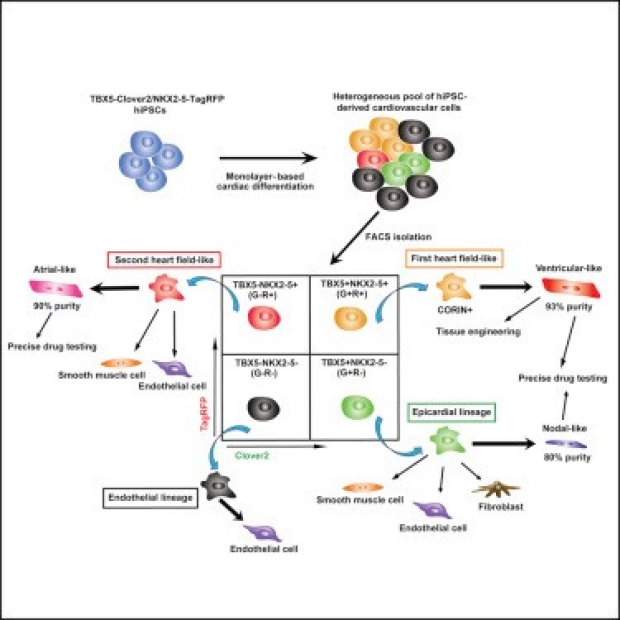
Figure: Four distinct hiPSC-CM lineages are isolated using the newly-developed double reporter tool. Lineage-specific cardiomyocytes can be used for precise drug testing.