Purifying Stem Cells of Heart Muscle
By Adrienne Mueller, PhD
February 25, 2021
Stem cells are powerful tools for understanding physiology and disease. Researchers often use induced pluripotent stem cells (iPSCs) in their studies. "Induced" means that the stem cells are created, or induced, from non-stem cells – usually fibroblasts collected by a simple blood draw. "Pluripotent" means the stem cells are able to become any different cell type: for example liver, lung, brain, or heart. Studies can therefore use human iPSCs (hiPSCs) as a minimally invasive, renewable, and versatile resource to study specific tissues and cell types. By creating heart muscle cells from hiPSCs, scientists are able to investigate heart cell function and a wide variety of cardiovascular diseases and therapies.
Heart muscle cells have different morphologies and properties depending on which chamber of the heart they are a part of. The heart has four chambers: two atria and two ventricles. Usually, when stem cells are used to derive cardiomyocytes, a heterogeneous mix of atrial and ventricular cardiomyocytes is the result. However, many diseases primarily involve exclusively atrial or ventricular cells. For example, hypertrophic or dilated cardiomyopathies and pulmonary hypertension are disorders that primarily involve the ventricles, whereas atrial fibrillation is specific to atrial cardiomyocytes. In order to understand diseases that primarily affect one chamber type, it is important to be able to study the heart cells that comprise that chamber in isolation—without contamination of other cell types.
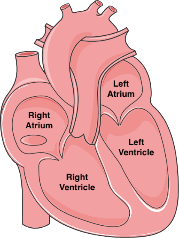
The four chambers of the human heart.
In a study recently published in Scientific Reports by first author Orlando Chirikian and senior author Sean M. Wu, MD, PhD, investigators developed a new method to isolate stem cell-derived atrial and ventricular heart muscle cells. To label atrial cardiomyocytes, they used CRISPR-Cas9 technology to insert a small piece of DNA that codes for a blue fluorescent protein into the genomes of stem cell next to sarcolipin, an atrial-specific gene. Similarly, the DNA for a red fluorescent protein, was inserted adjacent to a ventricular-specific gene. By inserting DNA for these two differently-colored reporters, the investigators could ensure that blue fluorescent protein would only be present in atrial cells and the red fluorescent protein only in ventricular cells. The investigators were then able separate fluorescent blue atrial cells from red ventricular cells thereby creating purified populations of cardiomyocytes for further experiments. By developing novel methods to purify stem cell-derived heart muscle cells, this study has created a platform for future work to improve our understanding of atrial- and ventricular-specific diseases and help us develop new therapies.
Other Stanford Cardiovascular Institute-affiliated authors include William R. Goodyer, Elda Dzilic, Vahid Serpooshan, Jan W. Buikema, Wesley McKeithan, HaoDi Wu, Guang Li, Soah Lee, Markus Merk, Francisco Galdos, Aimee Beck, Alexandre J. S. Ribeiro, Sharon Paige, Mark Mercola, Joseph C. Wu, and Beth L. Pruitt.

Dr. Sean Wu